If you’ve ever stepped into a cardiology clinic and seen a team huddled around a screen where the heart’s rhythmic, beating shadow lights up in crisp detail, chances are you’ve glimpsed cardiovascular ultrasound at work. As the owner of a cardiovascular ultrasound supply business, I get asked this question all the time: Can cardiovascular ultrasound actually detect heart valve diseases? The short answer is a resounding yes—but let’s dig into why this technology isn’t just a diagnostic tool, but the gold standard for catching valve issues before they become life-threatening. I’ve spent 12 years in this industry, helping clinics upgrade their equipment and training teams to use these tools, and I’ve seen firsthand how the right ultrasound machine can turn vague symptoms into clear, actionable answers. Let’s break this down, no jargon overload, just real talk about what this technology does. Cardiovascular Ultrasound

First, let’s get on the same page about what heart valve diseases are. The human heart has four valves—mitral, aortic, tricuspid, and pulmonic—each with a specific job: to let blood flow in one direction, keeping your circulatory system moving smoothly. When a valve goes wrong, it’s usually one of two ways: stenosis, where the valve becomes stiff and narrow, making it hard for blood to pass through, or regurgitation (sometimes called “leaky valve”), where the valve doesn’t close all the way, so blood flows backward instead of forward. Early stages might cause nothing more than a little fatigue or shortness of breath when you exercise—symptoms that are easy to brush off as stress or age, especially for people in their 50s and 60s. That’s where cardiovascular ultrasound steps in; it’s non-invasive, it’s fast, and it can pick up issues no stethoscope can.
Let’s start with the most common type of heart valve disease: aortic stenosis. Over time, calcium can build up on the aortic valve, hardening the tiny flaps until they can’t open fully. A lot of patients come in saying they get winded walking up a flight of stairs, and their primary care doc might hear a faint murmur on auscultation. A stethoscope can’t tell you how bad that stenosis is, though. It can’t measure the pressure difference across the valve, or calculate the exact area the valve is letting blood through—that’s where echocardiography, the specific type of cardiovascular ultrasound used for the heart, comes in. My team and I supply high-resolution ultrasound machines that can do this with a simple transthoracic echocardiogram (TTE): a wand, a little gel, and a 30-minute scan of the chest. We can see the calcium deposits, watch how the valve opens and closes in real time, and get precise numbers that tell cardiologists if a patient needs medication, lifestyle adjustments, or even surgery. Last year, I worked with a small clinic in rural Ohio that had been missing aortic stenosis cases because their old ultrasound machine was too grainy to pick up subtle valve thickening. When we upgraded their equipment, they caught three moderate stenosis cases in patients who thought their fatigue was just part of getting older. That’s the difference good cardiovascular ultrasound makes.
Next up is mitral regurgitation, another super common valve issue—roughly 2.5 million people in the U.S. alone live with it, according to the American Heart Association. Mitral regurgitation happens when the mitral valve (between the left atrium and left ventricle) doesn’t seal properly, so blood flows back into the atrium every time the heart contracts. Mild cases might not cause symptoms for years, but severe regurgitation can lead to heart failure if it’s not caught early. The tricky part with mitral valve issues is that symptoms are often vague: occasional dizziness, heart palpitations, swollen ankles. A lot of doctors might order an X-ray, but an X-ray only shows the heart’s size—it can’t show the valve itself. Cardiovascular ultrasound does both. A TTE can visualize the mitral valve’s flaps, check if they’re prolapsing (bulging backward into the atrium) or if the chordae tendineae—those thin, string-like structures holding the valve in place—are stretched or damaged. For more detailed views, we also supply transesophageal echocardiograms (TEE), which use a small ultrasound wand inserted down the esophagus, right next to the heart, so the image is even clearer. TEE is often used before valve repair surgery, but it’s also great for catching subtle regurgitation that might not show up on a regular TTE. I remember a 62-year-old teacher I met at a trade show a few years ago; she’d been dealing with “brain fog” and random palpitations for months, and her doctor couldn’t figure out why. We walked her clinic through a TEE demonstration on a demo machine, and sure enough, she had mild mitral regurgitation that was causing small blood clots to form in her heart—no wonder she was foggy. That case stuck with me because it’s exactly why cardiovascular ultrasound is so critical: it doesn’t just listen to the heart—it shows it.
But wait, is cardiovascular ultrasound the only tool? No, and I’d never try to say that. Sometimes, for patients with larger body habitus or certain anatomical quirks, a TTE might not give the clearest view, and a TEE is needed. For some rare valve diseases, like infectious endocarditis (where bacteria form on the valve), we might pair ultrasound with a CT scan or MRI to get a full picture. But here’s what I always tell clinic directors: if you’re only using a stethoscope and basic blood tests to screen for valve disease, you’re missing cases. Blood tests can show inflammation, but they can’t show how the valve is functioning. Stethoscopes catch many murmurs, but they can’t distinguish between a harmless murmur and a murmur caused by a stenosis that’s already at a dangerous stage. Cardiovascular ultrasound is the only tool that gives you real-time, functional data about the valves—how they move, how much blood is flowing through them, if there’s any backflow. It’s the foundation of valve disease diagnosis.
Let me talk about something a lot of people don’t mention: accessibility. For decades, echocardiography was tied to large, bulky machines that only big hospitals could afford. But that’s changed a lot in the last 10 years, and as a supplier, I’ve been proud to bring high-quality, portable cardiovascular ultrasound machines to smaller clinics and urgent care centers. A lot of rural and community clinics were stuck relying on outside referrals for echo tests, which meant patients waited weeks to get answers. Now, with a portable machine that’s easy to operate, a clinic can do an echo on-site in 30 minutes, get results the same day, and start treatment faster. Last year, I worked with a clinic in West Virginia that used to send all their cardiac patients 2 hours away for echo scans. Now, they have a portable machine from our line, and they’ve cut wait times for valve disease screening from 14 days to 24 hours. That’s not just good for business—it’s good for patients. Catching valve disease early means less invasive treatment, lower healthcare costs, and better outcomes.
Now, let’s address a common misconception: some people think cardiovascular ultrasound is only for diagnosing existing disease, not for monitoring it. But that’s not true at all. Once a patient is diagnosed with a mild valve issue, their cardiologist will order regular echo scans to track how the valve is changing. For example, a patient with mild aortic stenosis might get an echo every year to see if the calcium buildup is getting worse, or if the stenosis is progressing to moderate or severe. This monitoring is crucial because severe valve disease often requires surgery, and catching that before symptoms get bad is key. I’ve had clients tell me that their patients with mild valve issues have been coming in for annual scans for 10 years, and the ultrasound has caught small changes that would have led to serious problems if they’d gone unnoticed. It’s like getting a checkup for your heart valves every year—way more precise than a regular physical.
I also want to mention the role of training here, because a machine is only as good as the person using it. When I supply a cardiovascular ultrasound machine to a clinic, I don’t just drop it off and leave. My team provides hands-on training for the techs and cardiologists, teaching them how to get clear images of all four valves, how to measure valve function, and how to spot subtle signs of disease. For example, it takes practice to tell the difference between a harmless heart murmur and a murmur from a leaking mitral valve, or to notice the early signs of tricuspid regurgitation, which is common in people with sleep apnea. I’ve seen clinics buy top-of-the-line machines but struggle because their staff didn’t know how to use them properly. That’s why my business doesn’t just sell equipment—we provide ongoing support, training, and regular maintenance to make sure our clients get the most out of their cardiovascular ultrasound tools.
Let’s get back to the original question: Can cardiovascular ultrasound detect heart valve diseases? If you’ve read this far, you already know the answer is a definite yes. It’s the primary diagnostic tool for every type of valve disease, from mild stenosis to severe regurgitation, from congenital valve issues (present at birth) to age-related or disease-related damage. It’s non-invasive, safe (no radiation, unlike CT scans), fast, and accessible now more than ever, thanks to portable models and smaller, user-friendly machines. I’ve seen it catch disease in patients who thought they were just tired, in athletes who were experiencing unexplained shortness of breath, in elderly patients whose symptoms were written off as old age. It’s not just a diagnostic tool—it’s a lifeline.
If you’re a clinic director, cardiologist, or healthcare administrator reading this, and you’re looking to upgrade your cardiovascular ultrasound capabilities, or if you’re starting a new cardiology practice and need reliable equipment, I’m here to help. We’ve worked with clinics of all sizes, from small rural practices to large urban hospitals, and we tailor our solutions to fit your needs and budget. Don’t wait until you miss a case of valve disease because your old equipment isn’t up to par. Reach out to our team to learn more about our cardiovascular ultrasound products, training, and support services.
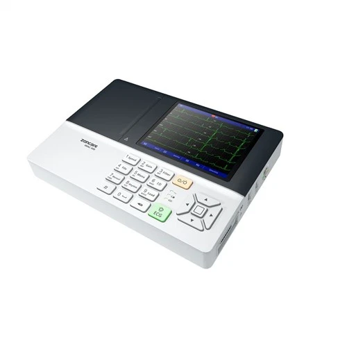
12 Lead ECG Machine References
- American Heart Association. (2023). Heart Valve Disease Statistics. Retrieved from American Heart Association database
- Otto, C. M., & Pearlman, A. S. (2021). Textbook of Clinical Echocardiography (7th ed.). Elsevier.
- Nishimura, R. A., et al. (2020). 2020 ACC/AHA Guideline for the Management of Patients With Valvular Heart Disease: A Report of the American College of Cardiology/American Heart Association Joint Committee on Clinical Practice Guidelines. Journal of the American College of Cardiology, 76(14), e125–e200.
- Zoghbi, W. A., et al. (2019). Recommendations for Noninvasive Evaluation of Native Valvular Heart Disease: A Report from the American Society of Echocardiography. Journal of the American Society of Echocardiography, 32(1), 1–56.
Wuhan Zoncare Bio-medical Electronics Co., Ltd.
Wuhan Zoncare Bio-medical Electronics Co., Ltd. is one of the leading cardiovascular ultrasound manufacturers and suppliers in China. We warmly welcome you to wholesale discount cardiovascular ultrasound from our factory. All customized products are with high quality and low price. Contact us for more cheap products.
Address: Zoncare building, No. 380, High-tech 2nd Road, Eastlake High-tech Development Zone, Wuhan, Hubei 430206 P. R. China.
E-mail: info@zoncare.com
WebSite: https://www.zoncareglobal.com/
